Welcome to Hull York Medical School
Continuing professional development

Radiological Anatomy

Course length

12 weeks part-time

Start date

February 2027

Location

Hull campus

Apply now
Health Technology and Human Anatomy students

Build advanced knowledge of radiological anatomy through an immersive combination of interactive lectures, guided practical sessions and independent study. 

This short course is designed for those who want to develop a rigorous, clinically contextualised understanding of normal and variant anatomy as seen through the lens of medical imaging. Through detailed exploration of commonly used radiological modalities, you will strengthen your ability to interpret anatomical structures with precision and apply that knowledge confidently in clinical, educational or research settings.

A distinctive feature of this course is its integration of flipped learning with hands-on practical sessions using a standardised bank of anonymised radiological images, accessed through standard image-viewing software.

Each learning week builds sequentially on the last, enabling you to develop a comprehensive spatial understanding of the radiological anatomy of the entire human form. Lectures provided by radiologists help signpost the relevance and transferability of key knowledge, skills and principles across clinical and specialist contexts.

Key areas include:

  • Physics and practicalities of X-ray (radiographs and CT), MRI and ultrasound imaging
  • Regional radiological anatomy of the musculoskeletal system, thorax, breast, abdomen, pelvis, head and neck, and central nervous system
  • Systematic radiological anatomy of the genitourinary, gastrointestinal and vascular systems
  • Normal and common variant anatomy in routinely used radiological modalities
  • Live anatomy using ultrasound, including image acquisition and optimisation
  • Interpretation of radiological images using real anonymised patient data

Learning activities include:

  • Flipped interactive lectures built on preparatory video, radiographic and textual teaching material
  • Guided practical sessions using an anonymised image bank and standard image-viewing software
  • Live anatomy sessions using ultrasound devices to explore three-dimensional anatomy in two-dimensional form
  • Structured practical workbook tasks including outlining, segmenting and identifying defined anatomical structures
  • Self-directed learning, self-assessment materials and tutor support through the virtual learning environment
  • Onsite and offsite access to an image bank for independent study and workbook completion

You will receive 20 Level 7 credits on completing this short course, including passing the assessments.

Contact Us

[email protected] 01904 321690
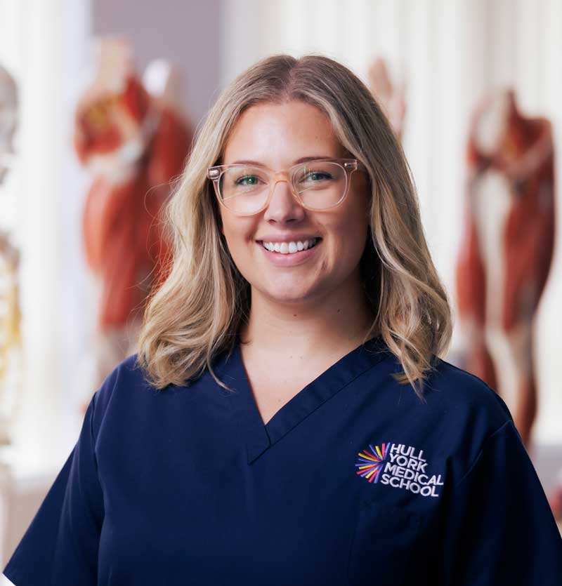
Lauren Clunie
Dr Lauren Clunie, Senior Lecturer in Clinical AnatomyView profile
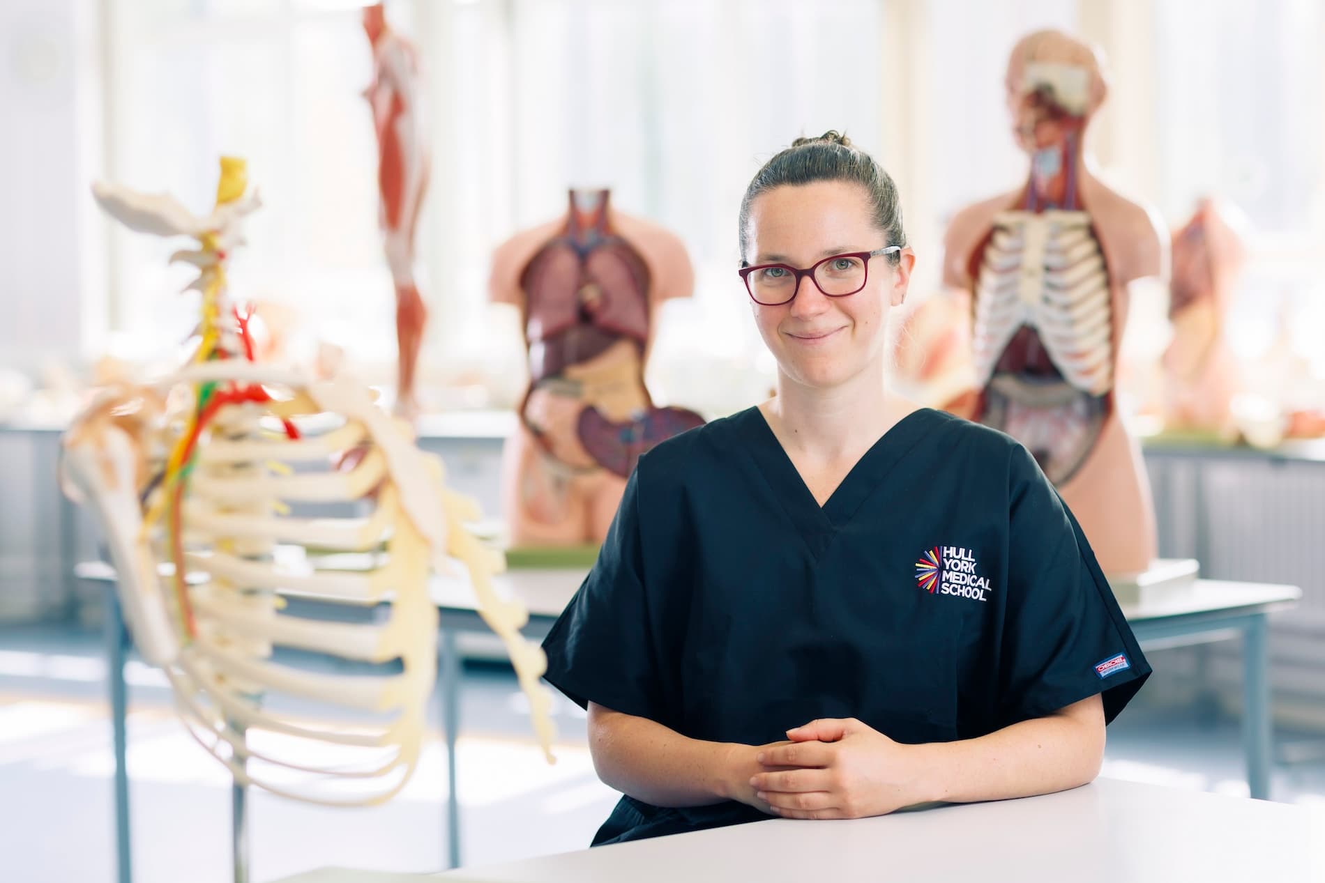
Dr Emily Hunter
Dr Emily Hunter, Lecturer in Clinical AnatomyView profile
University of Hull - Hull York Medical School

Who is this short course for?

This course is suited to healthcare professionals, clinicians in training, anatomy educators, radiographers, allied health professionals and researchers who want to strengthen their applied understanding of radiological anatomy and medical imaging interpretation. It is also appropriate for those wishing to develop skills in anatomical image analysis for professional practice, specialist development, teaching or further study in medical sciences.

Eligible applicants may take this course as a standalone CPD option or as part of the PGCert/PGDip/MSc in Clinical Anatomy and Education or PGCert/PGDip/MSc in Clinical Anatomy.

Teaching and assessment

What you will learn

You will study the normal and variant anatomy of the human body as visualised through commonly used clinical imaging modalities, including X-ray, CT, MRI and ultrasound. The course explores the physics underpinning each modality, the practicalities of imaging different body regions, and the interpretation of regional and systematic radiological anatomy using real anonymised patient data. Practical workbook tasks and live ultrasound sessions develop technical skills alongside anatomical knowledge, building a comprehensive spatial understanding of the radiological human form.

By the end of this course, you will be able to:

  • Describe the physics of image creation using X-ray, CT, MRI and ultrasound, and discuss the relative strengths and limitations of each modality.
  • Describe the bony and soft tissue anatomy visible on radiographs and fluoroscopic studies, including common and clinically significant variants.
  • Describe the radiological anatomy visible on CT and MRI across the major body regions and systems.
  • Generate and optimise ultrasound images of commonly scanned muscles and tendons, applying skills of medical image interpretation to the study of clinical anatomy.
  • Critically analyse anatomical variation in radiological imaging and integrate observations to construct a greater comprehension of the complexity of the human body in clinical practice.

How is this short course delivered?

The course is delivered through a series of flipped interactive workshops and guest seminars from clinicians.

Preparatory material, including video, radiographic and textual content covering imaging physics, regional anatomy and practical imaging considerations will be provided through the virtual learning environment ahead of each taught session. Interactive lectures then build on this foundation, applying background knowledge to the interpretation of normal, variant and pathological anatomy using real anonymised patient data.

Practical sessions provide you with a standardised bank of anonymised radiological images, accessed using standard image-viewing software, through which you complete structured tasks including outlining, segmenting and identifying defined anatomical structures.

Live anatomy sessions using ultrasound devices further develop your appreciation of three-dimensional anatomy in two-dimensional form, as well as your skills in acquiring clinically relevant images.

Direct tutor interaction throughout the course enables you to build on your learning and tailor your study to your own professional, academic or specialist interests.

Structured learning resources and tutor support are available through the virtual learning environment, with both onsite and offsite access to the image bank to support flexible independent study.

Time commitment

As a 20 credit level 7 module, the total time commitment, including contact time and self-directed learning, is 200 hours. Across the module, this is approximately 16 hours per week, including timetabled sessions.

This time allocation should include a minimum of 80 hours of teaching practice, including planning, preparation and assessment.

Assessment

Assessment is centred on a summative radiological workbook (3,000 words, 80%) in which you evidence your practical skills and anatomical knowledge through structured tasks completed across the module.

Formative assessment comprises a section of the workbook submitted mid-trimester for tutor feedback before the final submission.

The module also includes a summative group oral presentation delivered mid-trimester, contributing 20% to the final module mark.
Applying

Entry requirements

An undergraduate (strong 2.2 or above) or postgraduate degree in a relevant subject containing significant anatomy, such as:

  • Anatomy, Medicine, Medical Engineering, Physician Associate Studies, Veterinary Medicine
  • Biological or Biomedical Science and related programmes (Zoology, Bioengineering), with significant evidence of anatomy study (curricular or extra-curricular)

Applicants are encouraged to highlight previous anatomical experience in the application. 

Fees and funding

Tuition Fees

The fee for this short course for the academic year 2026/27 is

  • Home (UK): £1,494 
  • International (including EU): £3,650

Fees are reviewed annually and may change.

University of Hull - Hull York Medical School

applying

How to apply

Applications for this short course will be accepted soon. Sign up for our Continuing Professional Development newsletter to be informed when applications open.

The deadline for applications is 9 January 2027.

If you have any enquiries, please email [email protected].

Get our continuing professional development newsletter Subscribe now